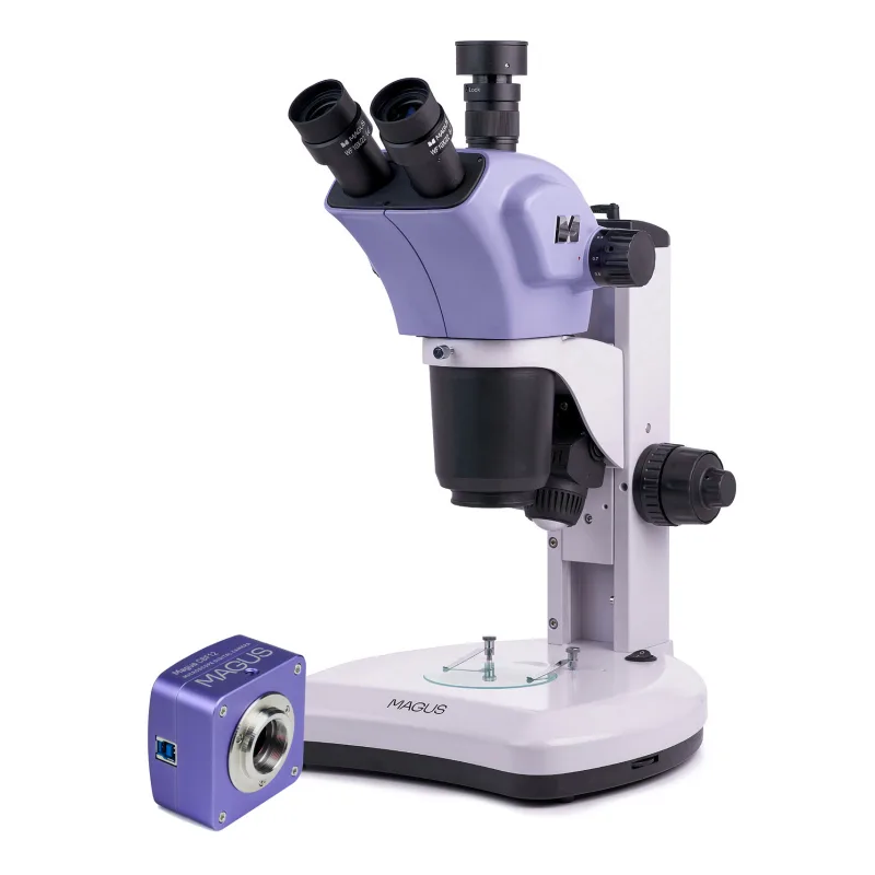
MAGUS Stereo D7T PLUS Digital Stereomicroscope
MAGUS Stereo D7T PLUS Digital Stereomicroscope GPSR: Levenhuk Europe V Chotejně 700/7 102 00 Praha 10 Česká republika 📞 +420 246 019 025 🌐 eu.levenhuk.com Bezpečnostné upozornenia – mikroskopy…
Specifikacia MAGUS Stereo D7T PLUS Digital Stereomicroscope
MAGUS Stereo D7T PLUS Digital Stereomicroscope
GPSR: Levenhuk Europe V Chotejně 700/7 102 00 Praha 10 Česká republika 📞 +420 246 019 025 🌐 eu.levenhuk.com Bezpečnostné upozornenia – mikroskopy Mikroskop používajte iba podľa určenia a v súlade s návodom výrobcu. Pri modeloch s osvetlením (LED/halogén) nepozerajte priamo do zdroja svetla – môže dôjsť k poškodeniu zraku. Nevystavujte zariadenie vlhkosti, vode ani extrémnym teplotám – hrozí poškodenie alebo úraz elektrickým prúdom.
Používajte iba originálne napájacie adaptéry a príslušenstvo odporúčané výrobcom. Nikdy nesmerujte mikroskop (najmä so zrkadlom alebo silným svetlom) na Slnko – riziko trvalého poškodenia zraku. Pred čistením alebo údržbou odpojte zariadenie od elektrickej siete.
Manipulujte opatrne so sklenenými časťami (šošovky, preparáty) – riziko poranenia. Uchovávajte mimo dosahu malých detí bez dozoru – niektoré časti môžu byť malé alebo ostré. Pri práci s chemickými preparátmi dodržiavajte zásady bezpečnosti (rukavice, vetranie).
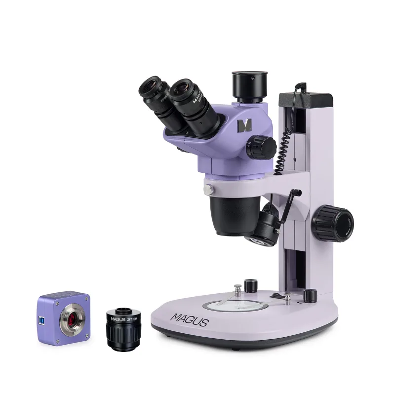
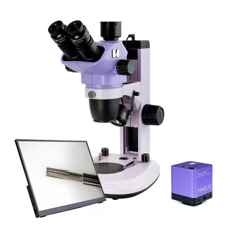
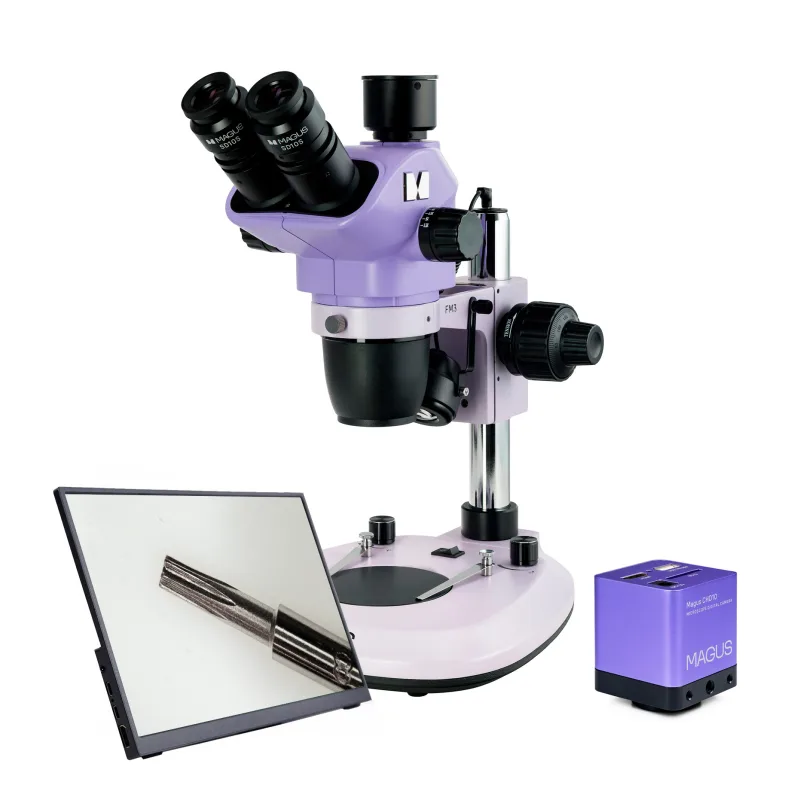
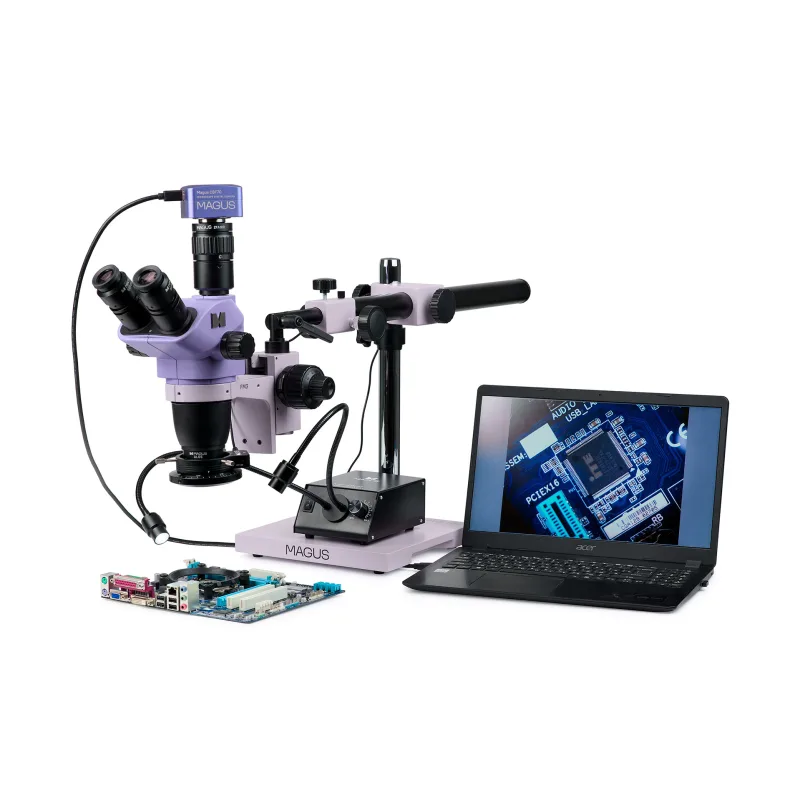
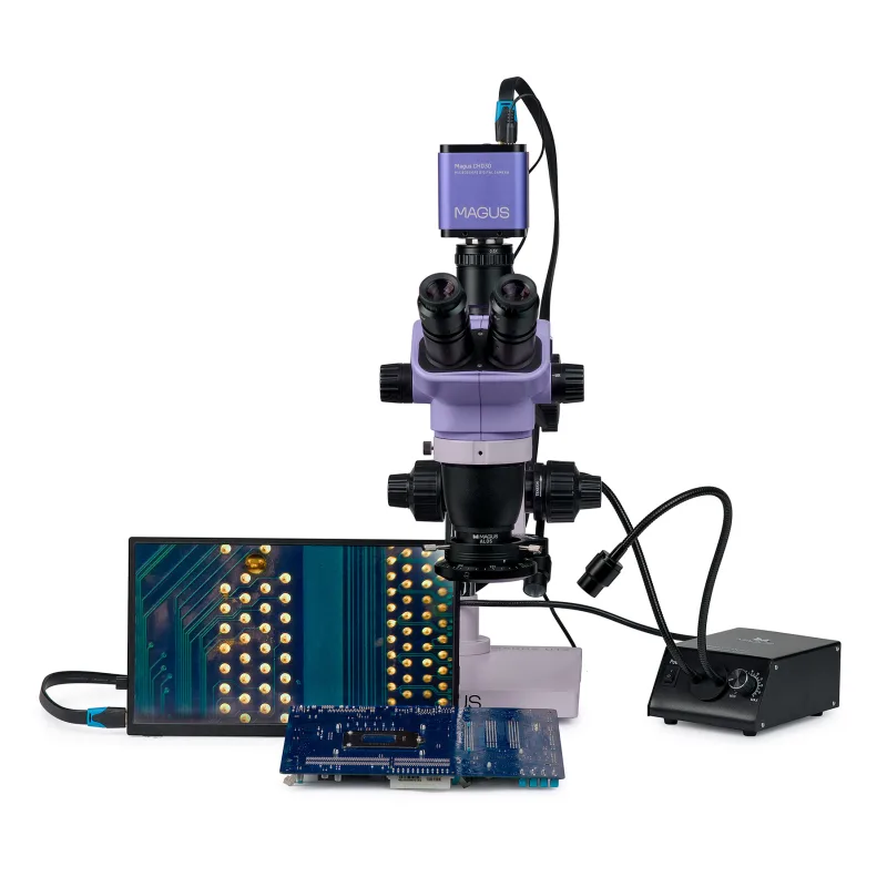
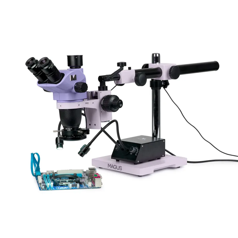
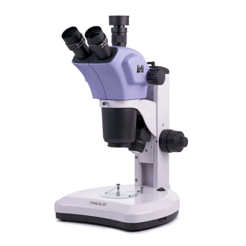
Zariadenie nerozoberajte – opravy prenechajte autorizovanému servisu.The stereomicroscope with the Greenough optical design provides accurate details of the surface topography with no loss of spatial orientation, ensuring a large depth of field when observing three-dimensional objects. The microscope smoothly changes magnification by a factor of 6.7 between 6.7x and 45x while maintaining a working distance of 105mm. It has built-in transmitted and reflected light illuminators for studying translucent and opaque objects.
The microscope provides for the installation of polarized light and darkfield accessories. The camera in the digital microscope allows the recording of the observation process as well as its intermediate and final outcomes. The 20MP sensor delivers the required resolution on the stereomicroscope.
The 1" sensor with a 1x adapter displays a field of view of 19.5x13.1mm on the computer screen with the microscope’s lowest magnification. The microscope is used for restoration, assembly, and quality control as well as in zoology and botany. Digital camera MAGUS CDF70 digital camera features low noise and high light sensitivity.
The camera is suitable for operation over the full magnification range of the microscope objective, ranging from 0.67x to 4.5x. The sensor size is 1". Therefore, a 1x C-mount adapter is used for mounting a camera on the microscope.
On such a sensor, the 1x adapter is intended to “show as large a field of view as possible with no distortion”. The 20MP sensor produces a realistic image in 5440x3648px. At this resolution, the frame rate is 20fps, which is enough to fine-tune the focus of the microscope.
At resolutions of 2736x1824px and 1824x1216px, the rate increases to 48fps and 58fps, respectively. Transitions between frames become smoother and the camera captures small movements of the object. These modes are suitable for classroom demonstrations, video recording, and observing moving objects.
The USB 3.0 provides 10 times faster data transfer rates than USB 2.0. Microscope head The trinocular head is 360° rotatable and can be locked in any desired position. The digital camera is mounted in the trinocular tube using a C-mount adapter.
The light path does not change. The splitting ratio is fixed: 80% to an eyepiece and 20% to the trinocular tube. The user observes the image in the eyepieces and on the screen at the same time.
The basic configuration includes 10x/22mm eyepieces with diopter adjustment and eye relief to work with glasses. Zoom objective The objective allows for the smooth change of magnification up to 6.7 times with no loss of focus while maintaining a large working distance of 105mm. The microscope generates an upright (not inverted) three-dimensional image.
Auxiliary objective lenses change the magnification range, field of view, and working distance of the microscope. Focusing mechanism Coarse focusing knobs are located on both sides of the stand. The coarse focusing tension is adjustable.
Stage plate A frosted glass or black-and-white plate is selected depending on the specimen. The frosted glass plate ensures even illumination and optimal light scattering when transparent and translucent objects are observed. The black-and-white plate is selected for observing opaque objects against a contrasting background: the black side for light objects and the white side for dark objects.
Light sources The microscope has a 3W LED transmitted light illuminator and a 3W LED reflected light oblique illuminator. The LED lifetime is 50,000 hours. Accessories There is a wide range of accessories for the microscope.
Eyepieces and auxiliary objective lenses extend the magnification range of the microscope. Digital cameras output images from the microscope
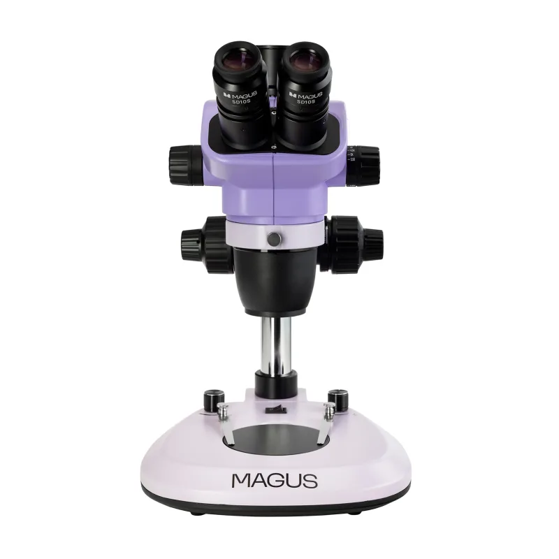
Stereomikroskop MAGUS Stereo 8T
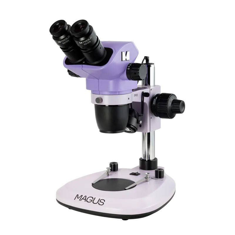
Stereomikroskop MAGUS Stereo 8B
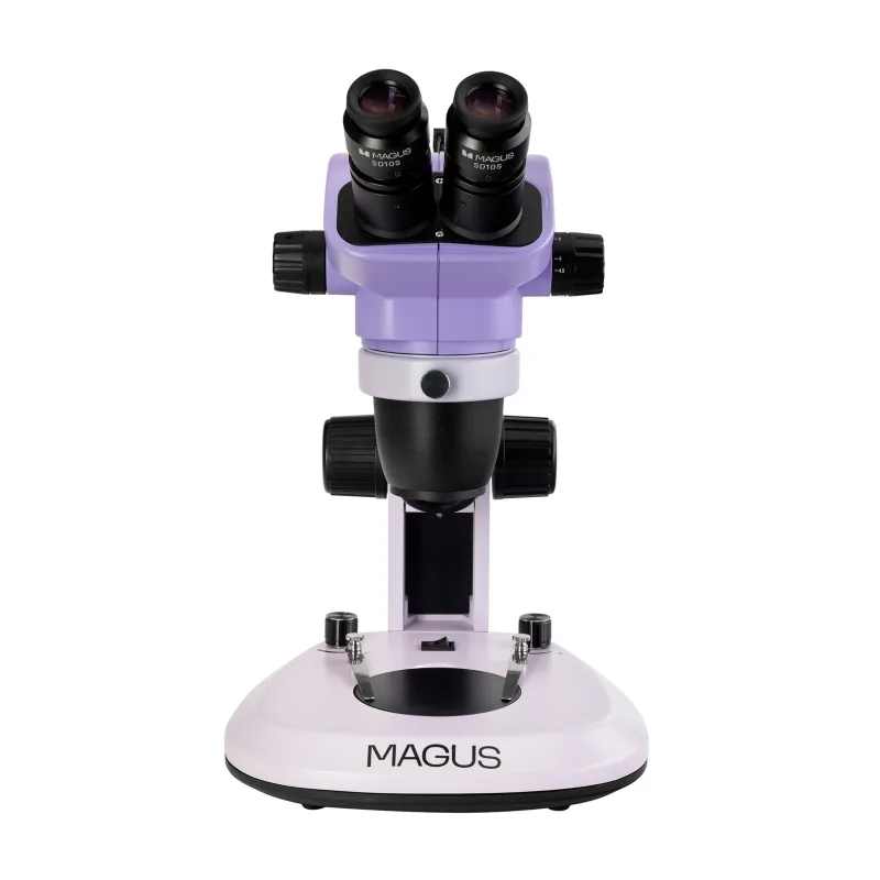
Stereomikroskop MAGUS Stereo 7B
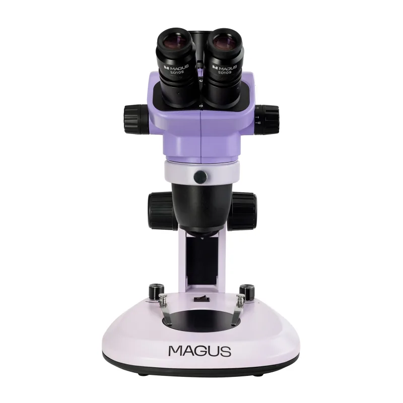
Stereomikroskop MAGUS Stereo 7T
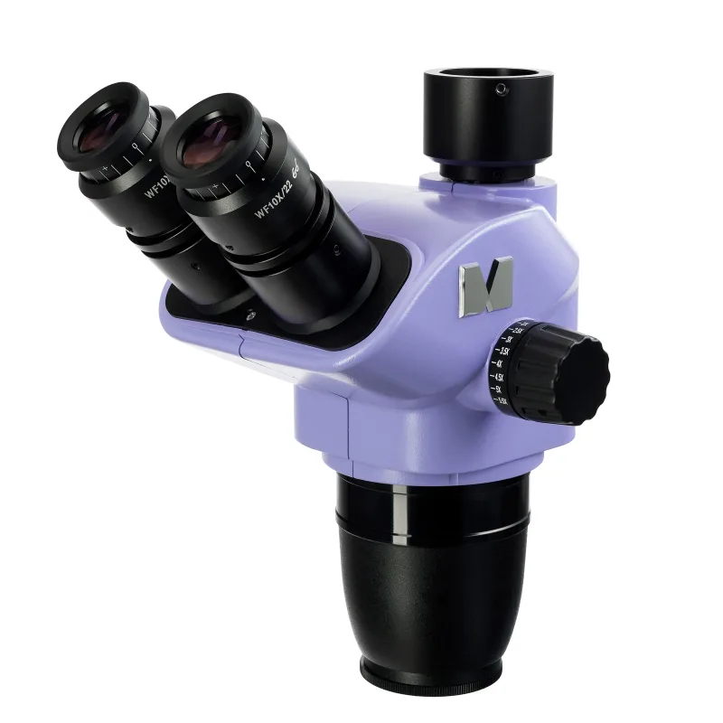
Hlavica mikroskopu MAGUS Stereo 8TH
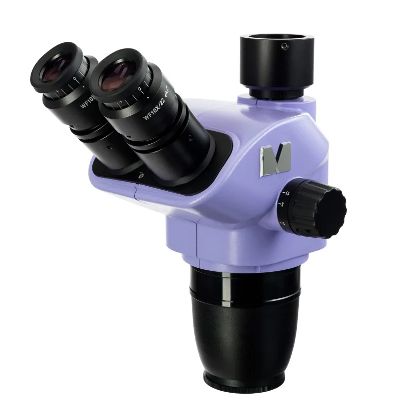
Hlavica mikroskopu MAGUS Stereo 7TH
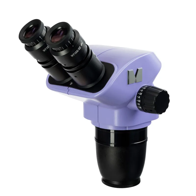
Hlavica mikroskopu MAGUS Stereo 8BH
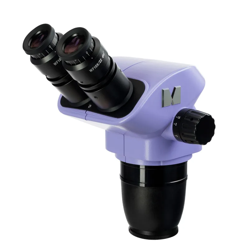
Hlavica mikroskopu MAGUS Stereo 7BH
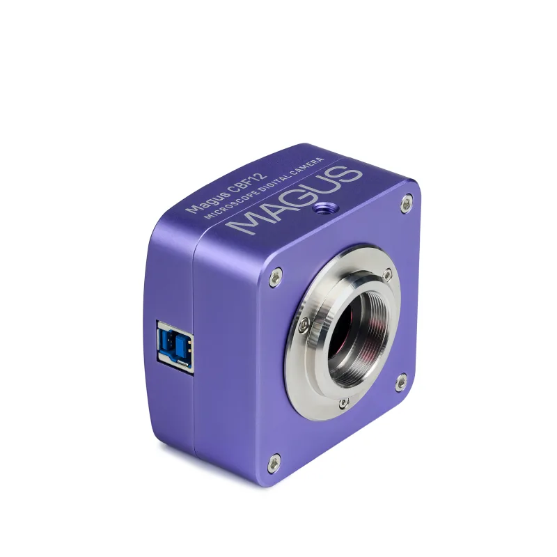
MAGUS CBF12 Digital Camera