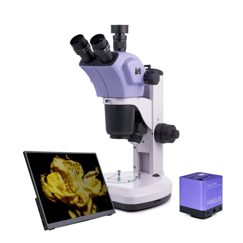
Digitálny Stereomikroskop MAGUS D9T LCD
Digitálny Stereomikroskop MAGUS D9T LCD GPSR: Levenhuk Europe V Chotejně 700/7 102 00 Praha 10 Česká republika 📞 +420 246 019 025 🌐 eu.levenhuk.com Bezpečnostné upozornenia – mikroskopy Mikroskop…
Specifikacia Digitálny Stereomikroskop MAGUS D9T LCD
Digitálny Stereomikroskop MAGUS D9T LCD
GPSR: Levenhuk Europe V Chotejně 700/7 102 00 Praha 10 Česká republika 📞 +420 246 019 025 🌐 eu.levenhuk.com Bezpečnostné upozornenia – mikroskopy Mikroskop používajte iba podľa určenia a v súlade s návodom výrobcu. Pri modeloch s osvetlením (LED/halogén) nepozerajte priamo do zdroja svetla – môže dôjsť k poškodeniu zraku. Nevystavujte zariadenie vlhkosti, vode ani extrémnym teplotám – hrozí poškodenie alebo úraz elektrickým prúdom.
Používajte iba originálne napájacie adaptéry a príslušenstvo odporúčané výrobcom. Nikdy nesmerujte mikroskop (najmä so zrkadlom alebo silným svetlom) na Slnko – riziko trvalého poškodenia zraku. Pred čistením alebo údržbou odpojte zariadenie od elektrickej siete.
Manipulujte opatrne so sklenenými časťami (šošovky, preparáty) – riziko poranenia. Uchovávajte mimo dosahu malých detí bez dozoru – niektoré časti môžu byť malé alebo ostré. Pri práci s chemickými preparátmi dodržiavajte zásady bezpečnosti (rukavice, vetranie).
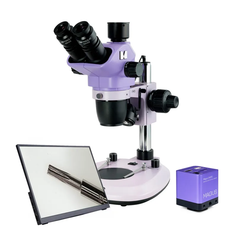
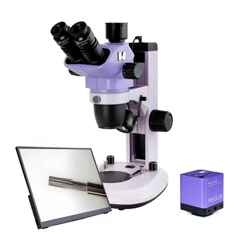
Zariadenie nerozoberajte – opravy prenechajte autorizovanému servisu.The MAGUS Stereo D9T LCD Stereomicroscope forms a non-inverted three-dimensional image and has a large working distance. You can examine samples but also conduct experiments with them. The Greenough optical scheme produces images that are sharp and three-dimensional which is essential for high-precision observations.
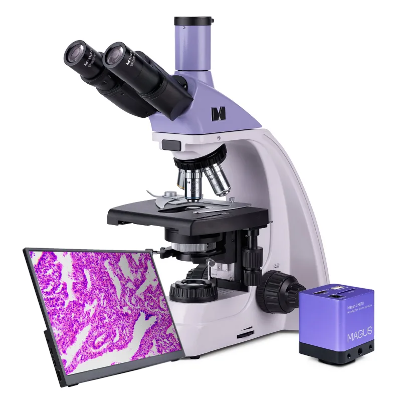
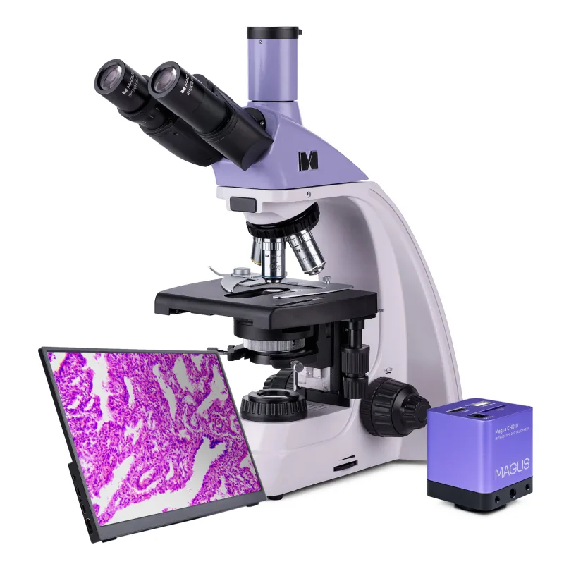
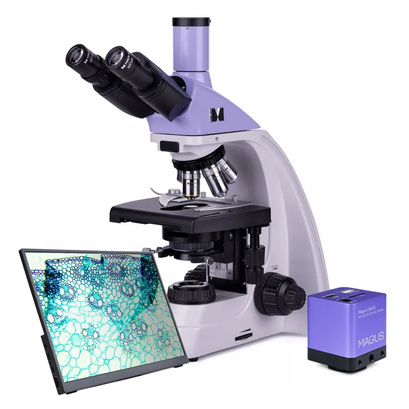
The microscope is equipped with a zoom lens and two illuminators (transmitted and reflected light) and can be optionally upgraded with additional accessories. Excellent choice for restoration, soldering, modeling, machinery service repair, and quality control. Microscope in use » Digital camera The digital HDMI-camera MAGUS CHD10 is equipped with a 2MP sensor and produces realistic Full HD images at a resolution of 1920x1080px.
It is an autonomous camera that does not require connection to a computer or the installation of additional software. The camera can be connected directly to a TV, monitor, or projector to display an image. The HDMI interface provides a high and stable transfer rate from the camera to the external screen.
Video is recorded at 60fps. The camera combines high FPS and high bandwidth HDMI. Therefore, videos are vivid, with no freezes or gaps between frames.
At maximum resolution, the image is well-detailed, moving objects are visible without bugs, and object movement is displayed without delays. Monitor MAGUS MCD monitors work in combination with MAGUS CHD cameras to form a complete imaging system for MAGUS microscopes. The connection to the camera occurs via its HDMI interface.
The monitor displays real-time images. Its IPS LCD display has a wide viewing angle, high brightness, and good contrast. When looking at the screen even from a large angle, the image is not distorted.
The screen size is 13.3''. The monitor can be installed on a horizontal surface using a folding stand or mounted directly on a camera or tripod. Optics The microscope has a trinocular head with a trinocular tube for mounting a camera.
A C-mount adapter is included, but a digital camera must be purchased separately. The trinocular head rotates 360°. The zoom objective combined with a 10x eyepiece provides magnification from 7x to 63x.
Thanks to the zoom lens, the magnification changes without reducing the focus. If you use auxiliary objectives (not included), you can change the magnification range, working distance, and field of view of the microscope. With optional accessories, the magnification of the microscope can be increased up to 315x.
Illumination The working area is lit by two LED illuminators: 5W transmitted light illuminator and 3W reflected light (oblique) illuminator. Both illuminators are AC powered, bright, do not change color temperature when adjusting brightness, and have a 50,000-hour life. Stage and focusing mechanism One of the included plates is installed on the stage: double-sided black and white, white, or transparent.
The first plate is used for observing opaque objects and makes the image high contrast. Dark-colored samples are examined on the white side, while light-colored samples - on the black side. White and transparent plates are used when you are observing translucent and transparent samples and want to achieve maximum brightness and uniform illumination of the working area.
The microscope has a coarse and fine focusing mechanism. Fine focusing is used at magnifications above 40x. Coarse focusing has tension adjustment.
Accessories To extend the microscope’s capabilities, use additional accessories such as eyepieces, auxiliary lenses, calibration slides, and digital cameras. It is also possible to install a device for polarized light observations. *Key features of the microscope:
Digitálny Stereomikroskop MAGUS D9T LCD
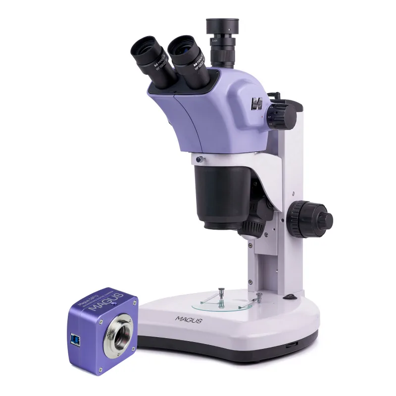
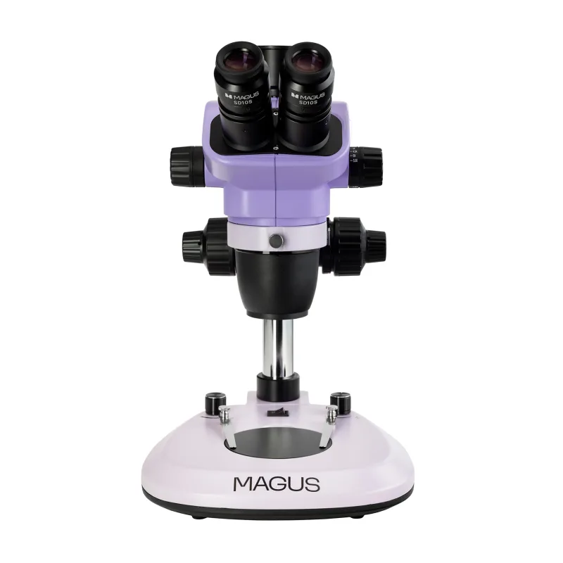
Stereomikroskop MAGUS Stereo 8T
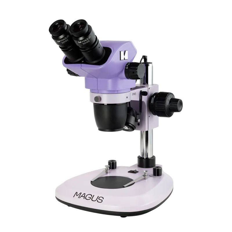
Stereomikroskop MAGUS Stereo 8B
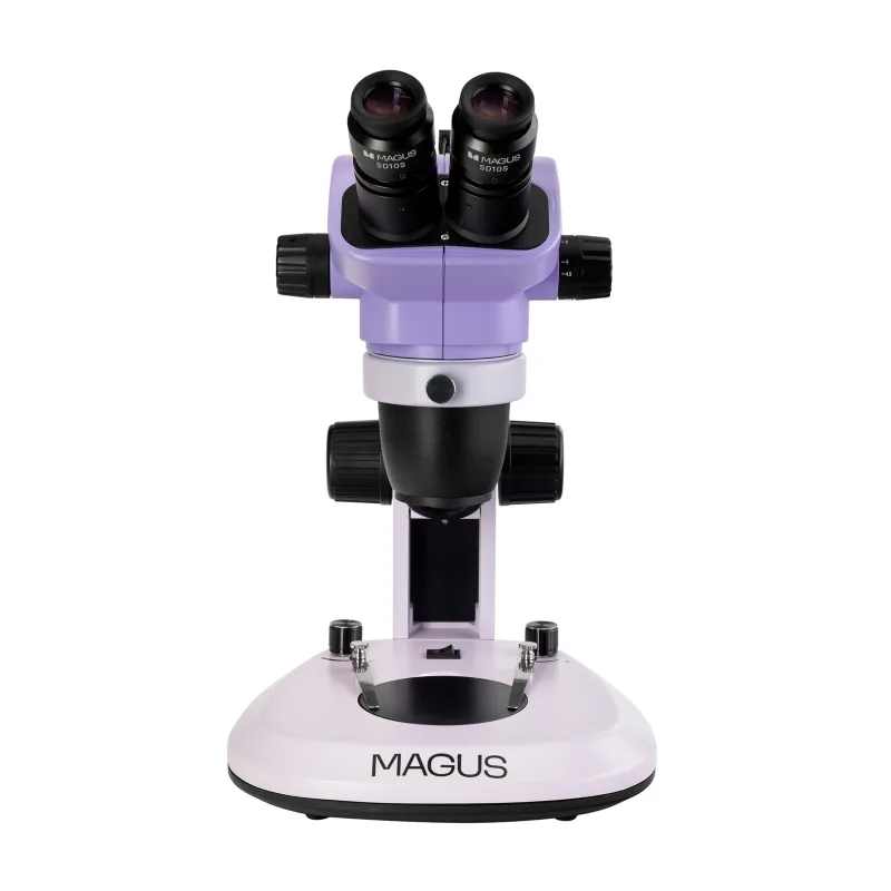
Stereomikroskop MAGUS Stereo 7B
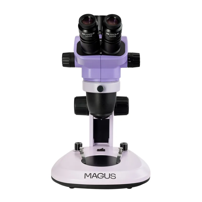
Stereomikroskop MAGUS Stereo 7T
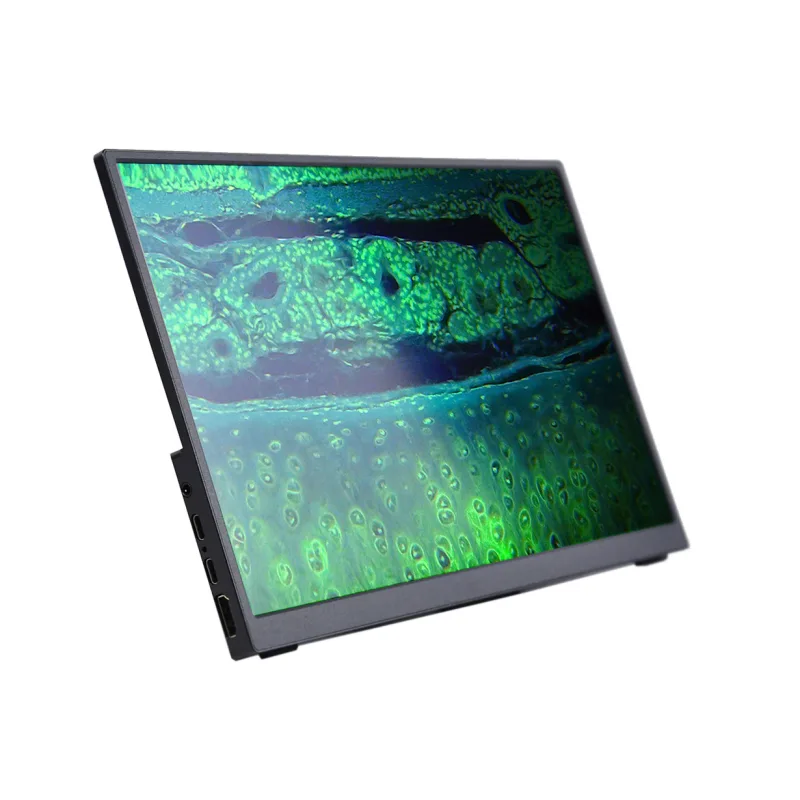
LCD monitor MAGUS MCD40
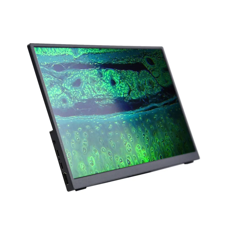
LCD monitor MAGUS MCD20

The Magus
The Magus
The Magus